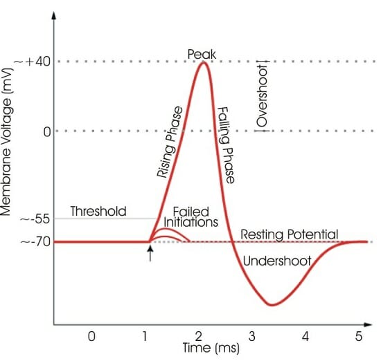

Voltage-gated sodium channels found in mammals can be divided into three types: Nav1.x, Nav2.x, and Nav3.x. A variety of alpha subunit voltage-gated sodium channels have been identified. Voltage-gated sodium channels can be divided into two subunits: alpha and beta. These channels are responsible for propagation of electrical signals in nerve cells. When electrically activated, they allow the movement of sodium ions across a plasma membrane. Voltage-gated sodium channels are proteins found in the membrane of neurons. G - glycosylation, P - phosphorylation, S - ion selectivity, I - inactivation, positive (+) charges in S4 are important for transmembrane voltage sensing. Voltage-Gated Channels Voltage-Gated Sodium Channel ĭiagram of a voltage-sensitive sodium channel α-subunit. Local voltage thresholds for dendritic spike initiation are usually higher than that of action potential initiation in the axon therefore, spike initiation usually requires a strong input. Dendritic spikes usually transmit signals at a much slower rate than axonal action potentials. Dendritic spikes can be generated through both sodium and calcium voltage-gated channels. If the voltage increases past a certain threshold, the sodium current activates other voltage-gated sodium channels transmitting a current along the dendrite. The influx of sodium ions causes an increase in voltage. Depolarization of the dendritic membrane causes sodium and potassium voltage-gated ion channels to open. They are one of the major factors in long-term potentiation.Ī dendritic spike is initiated in the same manner as that of an axonal action potential. Dendritic spikes have been recorded in numerous types of neurons in the brain and are thought to have great implications in neuronal communication, memory, and learning. Dendrites contain voltage-gated ion channels giving them the ability to generate action potentials. Alden Spencer, Eric Kandel, Rodolfo Llinás and coworkers in the 1960s and a large body of evidence now makes it clear that dendrites are active neuronal structures. However, the existence of dendritic spikes was proposed and demonstrated by W. Unlike its axon counterpart which can generate signals through action potentials, dendrites were believed to only have the ability to propagate electrical signals by physical means: changes in conductance, length, cross sectional area, etc. Dendritic signaling has traditionally been viewed as a passive mode of electrical signaling. They receive electrical signals emitted from projecting neurons and transfer these signals to the cell body, or soma. Dendrites are branched extensions of a neuron. In neurophysiology, a dendritic spike refers to an action potential generated in the dendrite of a neuron. Actual recordings of action potentials are often distorted compared to the schematic view because of variations in electrophysiological techniques used to make the recording. is a recording of an actual action potential N.B. shows the idealized phases of an action potential.


 0 kommentar(er)
